
Figure 2.
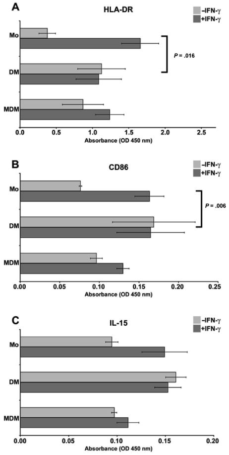
Analysis of activation markers on monocytes, purified decidual macrophages, and monocyte-derived macrophages using cell enzyme-linked immunosorbent assay (ELISA). Antibodies used to obtain the results were (A) anti-HLA-DR, (B) anti-CD86, and (C) anti-IL-15. In each panel, markers on blood monocytes (Mo), purified decidual macrophages (DM), and monocyte-derived macrophages (MDM) cultured in the absence (−IFN-γ) or presence (+IFN-γ) of 100 U/mL interferon-γ (IFN-γ) for 48 hours are shown. OD indicates optical density. Data shown are the means of values obtained in at least 3 separate experiments ± SEM. Brackets and P values indicate statistical significance between fold changes in response to IFN-γ treatment.