
Figure 4.
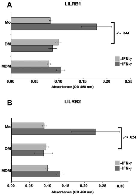
Analysis of HLA-G receptors on monocytes, purified decidual macrophages, and monocyte-derived macrophages by cell surface enzyme-linked immunosorbent assay. (A) Leukocyte immunoglobulin-like receptor (LILR)B1 and (B) LILRB2. Blood monocytes (Mo), purified decidual macrophages (DM), and monocyte-derived macrophages (MDM) were cultured in the absence (−IFN-γ) or presence (+IFN-γ) of 100 U/mL interferon-γ (IFN-γ) for 48 hours. OD indicates optical density. Data shown are the mean values obtained in 3 separate experiments ± SEM. Brackets and P values indicate statistical significance between fold changes in response to IFN-γ treatment.