
Figure 6.
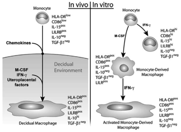
Schematic illustration showing marker expression of decidual macrophages (left panel) in comparison with in vitro–activated monocytes, differentiated monocytes, and monocytes that were both differentiated and activated (right panel). IFN, interferon; IL, interleukin; M-CSF, macrophage colony-stimulating factor; TGF, transforming growth factor.