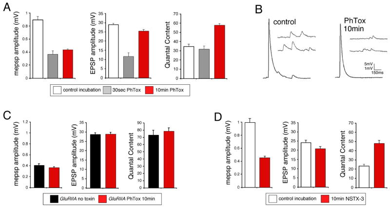
Figure 2. Rapid induction synaptic homeostasis in a semi-intact preparation.
A) Quantification of average mepsp amplitude (left), average EPSP amplitude (middle) and quantal content (right) for the three conditions listed below the graphs; 10min saline incubation (control incubation), 30 second incubation in PhTox (30sec PhTox) and a 10 minute incubation in PhTox (10min PhTox). There is a statistically significant increase in quantal content after a 10 min PhTox incubation (n=48; p<0.01) compared to a 30 sec PhTox incubation (n=10) or saline incubation (control incubation; n=42). B) Sample traces prior to and following PhTox incubation for 10min. Scale bar 150ms; 5mV for EPSPs and 150ms; 1mV for mepsps. C) Quantification as in (A). Incubating GluRIIA mutants for 10 min in PhTox (n=12) does not reduce average mepsp amplitude beyond that observed in GluRIIA mutants without toxin (p>0.2; n=12) and does not increase quantal content beyond that observed in GluRIIA mutants without toxin (p>0.5). D) Data are shown for wild-type synapses incubated in control saline (10 min) versus wild-type synapses incubated in NSTX-3 (NSTX-3; 10μM, 15 min). Prior to recording, synapses were washed in normal saline without NSTX-3. A persistent inhibition of postsynaptic receptors following the wash is shown by the significant decrease in mepsp amplitudes compared to mock treated wild-type controls (p<0.01). After a 15 min incubation in NSTX-3, EPSP amplitudes are near wild-type values (p>0.1) and there is a significant increase in quantal content (p<0.01).

