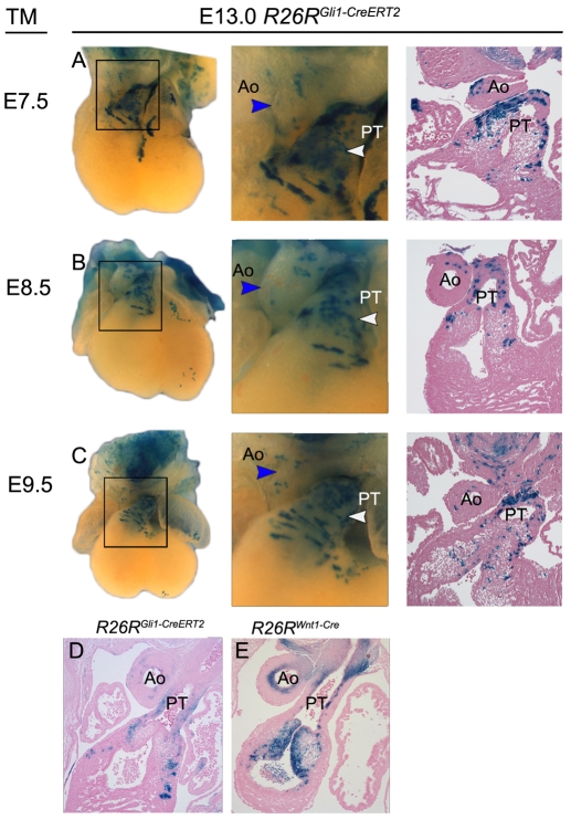
Fig. 2.
Hedgehog-receiving cells mark the pulmonary trunk. (A-C) Hh-receiving lineage marked in R26RGli1-CreERT2 embryos by a single dose of TM at E7.5 (A), E8.5 (B) or E9.5 (C), and analyzed at E13.0. β-Galactosidase positive cells identified in whole-mount (left and middle; 4× and 10× magnification, respectively) and cross-section (right, 10× magnification) histology. In the outflow tract, marked cells contribute specifically to the pulmonary trunk (white arrowheads and PT in A-E) and pulmonary trunk endocardial cushion at each time point. Few marked cells are observed in the aorta (middle, blue arrowheads; right, `Ao'). (D,E) Comparison of E13.5 outflow tracts marked by β-galactosidase expression in R26RWnt1-Cre (D) and R26RGli1-CreERT2 (E) mice. Cells that receive a Hh signal, marked by Gli1-CreERT2, primarily populate the outer edge of the outflow tract vessels, whereas cells originating from the neural crest, marked by Wnt1-Cre, primarily populate the inner wall of the outflow tract. Gli1-CreERT2 and Wnt1-Cre expressing cells appear to populate complementary domains that may overlap slightly, but not broadly.

