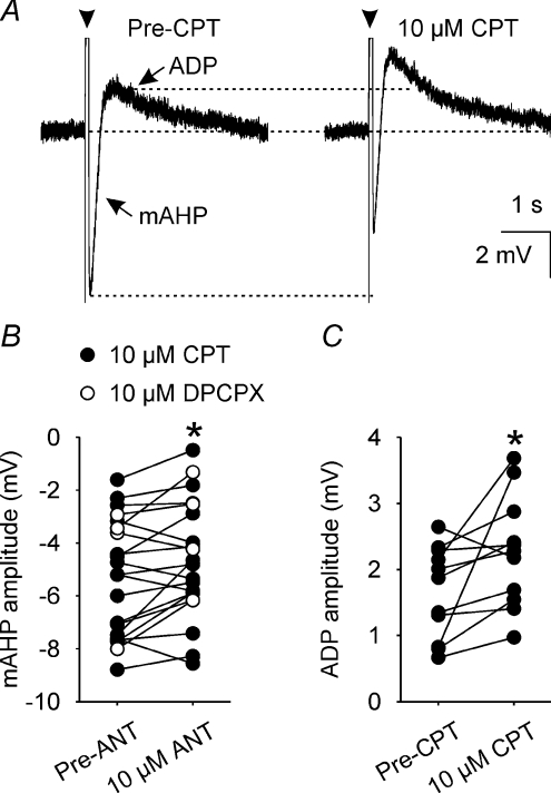
Figure 3. A1 receptor antagonism reduces mAHP amplitude.
A, mAHPs and ADPs (averages of 5 traces) that each follow a 5-spike train (arrowheads, spikes truncated) evoked by a 80 ms depolarizing pulse (+200 pA) before (Pre-CPT, left) and during superfusion of 10 μm CPT (right). B, mAHP amplitude in supraoptic nucleus neurones (n= 22) before (Pre-ANT, left) and during (10 μm ANT, right) superfusion of 10 μm of the A1 receptors antagonists CPT (•, n= 18) or DPCPX (○, n= 4), showing that A1 adenosine receptor antagonism reduces mAHP amplitude. *P < 0.05, two-way repeated measures ANOVA followed by Student–Newman–Keuls test. C, ADP amplitude in supraoptic nucleus neurones (n= 11) before (Pre-CPT, left) and during (right) superfusion of 10 μm CPT, showing that CPT increases ADP amplitude concomitant with its reduction of mAHP amplitude (note that none of the cells recorded in the presence of DPCPX displayed a measurable ADP). *P < 0.05, paired t test.

