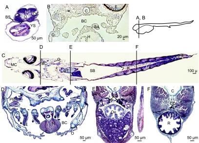
Fig. 2.
Histomorphology of the sea-bass at different stages, stained with Masson's trichrome (A,C–F) and in phase contrast (B), transverse sections (A,B,D–F) and longitudinal horizontal section (C). The location of transverse sections is indicated beside B and on C. Larvae at hatch (D0) (A), at D4 (B), and at D42 (C–F). At hatch, note the branchial slit (BS) and the straight digestive tract (DT) in an anterior section (A). At D4, the developing branchial cavity (BC) is limited by the developing opercula (O) and it contains the branchial arches (BA). At D42, the branchial cavity is well formed (C,D) with complete gills with arches (BA), filaments (F) and lamellae (L). In the median section (E), the urinary ducts (UD) are in a dorsal position compared to the oesophagus (Oe). In the posterior part of the body (F), the kidney is represented by the collecting ducts (CD) above the rectum (R). BA, branchial arch; BC, branchial cavity; BS, branchial slit; C, chord; CD, collecting duct; DT, digestive tract; E, eye; F, filament; Fi, pectoral fin; H, heart; I, ionocyte; Int, intestine; K, kidney; L: lamellae; M: muscle; MC: mouth cavity; NS: nervous system; O: operculum; Oe: oesophagus; OC: otic cavity; PB, pseudo-branches; PI, posterior intestine; R, rectum; RD, renal duct; SB, swim bladder; T, integument; TI, integumental ionocyte; UB, urinary bladder; UD, urinary duct; YS, yolk sac.

