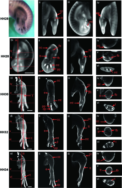
Fig. 4.
Expression of the Col XI gene in the chick hind limb at stages HH28 (a,b), HH30 (c), HH32 (d), HH34 (e). ai shows a dorsal view of a stage HH28 limb prior to scanning. External views of a volume representation of the same limb following OPT scanning and reconstruction are shown in aii (rotated 90º), aiii (rotated 180º) and aiv (rotated 270º). b–d (i) show similar external dorsal views of whole reconstructed limbs with red lines indicating the plane of virtual longitudinal (iii) and transverse (iv–vi) sections taken through the 3D reconstructions. Sections shown in b, c and d ii were taken parallel to the view shown in i. Expressing tissue appears as white/light grey in the reconstructions (noted in the joint capsule, early condensing cartilage, the perichondria and epiphyses of more mature condensations and connective tissues). cc, capsular condensation; pc, perichondrium; pp, proximal phalange; dp, distal phalange; fb, fibula; fe, femur; mt, metatarsal; tt, tibiotarsus; t, tendon; jl, joint line. Scale bar, 1 mm.

