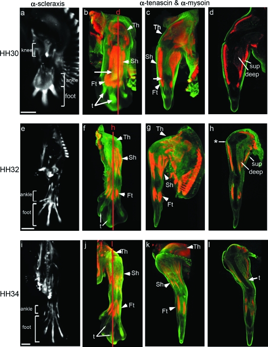
Fig. 5.
Development of the muscle and tendon system in the chick hind limb at stages HH30, HH32 and HH34. The left column shows limbs viewed from the dorsal aspect from whole 3D reconstructions showing the localization of Scleraxis transcripts (a,e,i) (in white). Remaining images show double labelled specimens using α-tenascin (green) and α-myosin (red) antibodies. The second and third columns show external views of whole specimens (dorsal, b,f,j; anterior, c,g,k). d, h and l are longitudinal sections indicated by red lines. Territories of knee, ankle and foot tendons are delimited by brackets. Th, thigh muscles; Sh, shank muscles; Ft, foot muscles; sup, superficial muscles; deep, deep muscles; t, tendons. * indicates the ilio-tibialis cranialis tendon. Arrow in c indicates residual connections between muscle blocks. Scale bars, 1 mm.

