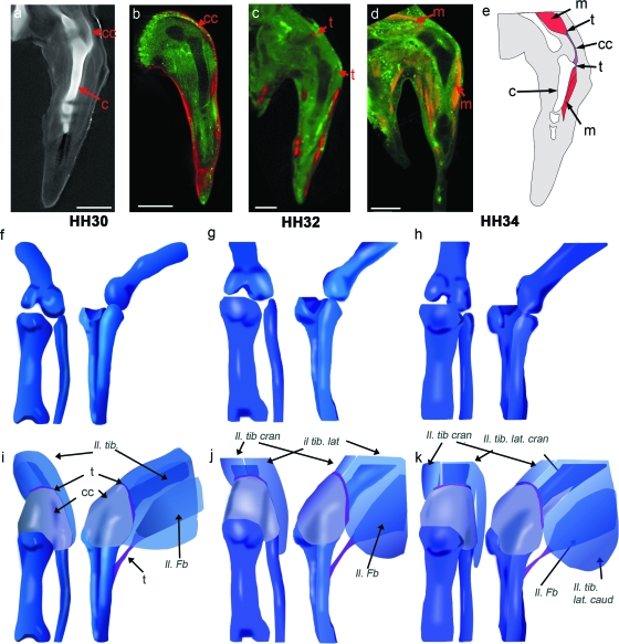
Fig. 6.
Building 3D representations of the chick knee joint at stages HH30, HH32 and HH34, incorporating multiple components. Specimens stained with Alcian blue (Fig. 6a) give the shape of the cartilaginous elements (Fig. 6e–h). a–d show sample comparable longitudinal sections through the mid tibiotarsus (HH32) of specimens stained with Alcian blue (a), in situ hybridized for Col XI (b, pseudocoloured in red) Scleraxis (c, pseudocoloured in red) and immunostained to reveal myosin (d). e shows integration of the data. For 3D representation of the integrated data, the developing cartilage anlagen (e–g) (Fig. 1) firms the base onto which the shape of the capsule, the various tendon attachment sites and associated muscles were added (full complement of tendons and muscle not shown) (i–k). Muscles (nomenclature as per Baumel et al (1979)): Il. Ti., ilio-tibialis (pre division into Il. tib. lat and Il. tib. cran); Il. tib. lat. (caud/cran), ilio-tibialis lateralis (caudalis/cranialis); Il. tib. cran., ilio-tibialis cranialis; Il. Fb., ilio-fibularis. c, cartilage; cc, capsular condensation; t, tendon; m, muscle. Scale bar, 1 mm.

