
Figure 4.
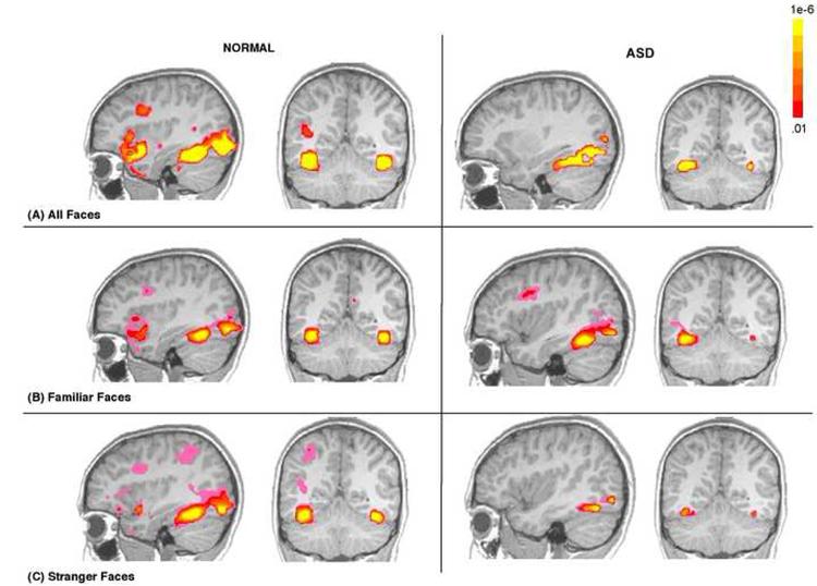
Functional activation maps illustrating the presence of significant functional activity in the fusiform in ASD and normal children in response to: (A) all faces combined (B), familiar faces only and (C) stranger faces only. While this figure highlights defects in fusiform function in response to stranger faces, it also illustrates that the fusiform is capable of functional responding in children with an ASD as depicted by robust functional activity in response to all faces combined and familiar faces. Data are shown at a voxel level of p<.01, overall alpha p<.05, whole brain corrected. The colors used in the functional maps represent p values associated with a t-statistic.