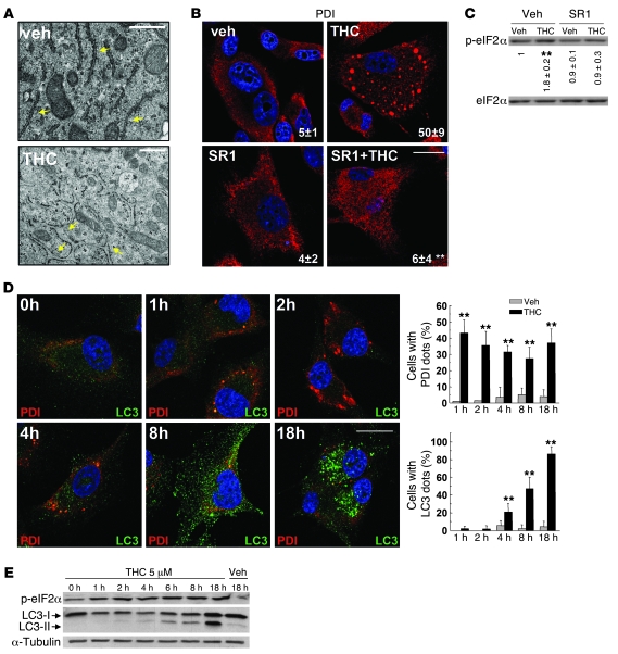
Figure 2. ER stress precedes autophagy in cannabinoid action.
(A) Effect of THC on U87MG cell morphology. Note the presence of the dilated ER in THC- but not vehicle-treated cells (6 h). Arrows point to the ER. Scale bars: 500 nm. (B) Effect of SR1 (1 μM) and THC on PDI immunostaining (red) in U87MG cells (8 h; n = 3). The percentage of cells with PDI dots relative to the total cell number is shown in the corner of each panel (mean ± SD). Scale bar: 20 μm. (C) Effect of SR1 (1 μM) on THC-induced eIF2α phosphorylation of U87MG cells (3 h; OD relative to vehicle-treated cells, mean ± SD; n = 3). (D) Effect of THC on PDI (red) and LC3 (green) immunostaining in U87MG cells (n = 3). The percentage of cells with PDI or LC3 dots relative to total cell number at each time point (mean ± SD) is shown. Scale bar: 20 μm. (E) Effect of THC on eIF2α phosphorylation and LC3 lipidation in U87MG cells (n = 3). **P < 0.01 compared with THC-treated (B) or vehicle-treated (C and D) cells.

