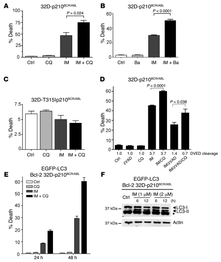
Figure 4. Inhibition of autophagy potentiates IM-induced cell death in 32D-p210BCR/ABL cells.
(A) 32D-p210BCR/ABL cells were cultured for 12 hours in the presence or absence of 1 μM IM, alone, or in combination with 5 μM CQ. Cell death was measured by annexin V staining. Values represent the mean ± SEM of 3 independent experiments. Data were analyzed by unpaired Student’s t test. (B) 32D-p210BCR/ABL cells were treated with 1 μM IM (12 hours), alone or in combination with 20 nM Ba. Cell death was measured as above. Values represent the mean ± SEM of 2 independent experiments. (C) CQ has no effects in 32D cells expressing the IM-resistant T315I p210BCR/ABL mutant. 32D cells expressing the IM-resistant T315I p210BCR/ABL mutant were treated with IM, with or without CQ, and analyzed for cell death induction as in A. (D) CQ-induced cell death is caspase independent. 32D-p210BCR/ABL cells were cultured for 6 hours in the presence or absence of different combination of 1 μM IM, 50 μM zVAD, and 5 μM CQ. The percentage of cell death was determined as above. Caspase activity was measured in extracts using a DVED fluorescent substrate. (E) Bcl-2 does not block CQ-mediated sensitization to IM. 32D-p210BCR/ABL cells ectopically expressing Bcl-2 (Bcl-2 32D-p210BCR/ABL) were cultured with 1 μM IM alone or in combination with 5 μM CQ. Cell death was measured as above. Values represent mean ± SEM (C–E). (F) Bcl-2 does not block IM-induced LC3-II accumulation. Western blot shows LC3-I/LC3-II levels in extracts of 32D-p210BCR/ABL cells ectopically expressing Bcl-2 treated for 6 or 12 hours with the indicated concentrations of IM. β-actin was detected as loading control.

