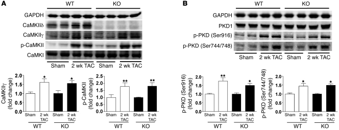
Figure 3. Upregulation of CaMKIIγ expression and PKD phosphorylation in cardiac hypertrophy.
(A) Expression of CaMKIIδ, CaMKIIγ, CaMKI, and phospho-CaMKII in LV homogenates 2 weeks after TAC, as detected by Western blot analysis. (B) Expression of PKD1 and phospho-PKD (Ser916 and Ser744/748) in LV homogenates 2 weeks after TAC were detected by Western blot analysis. Data are mean ± SEM of values from 4–6 determinations. *P < 0.05, **P < 0.01 versus sham.

