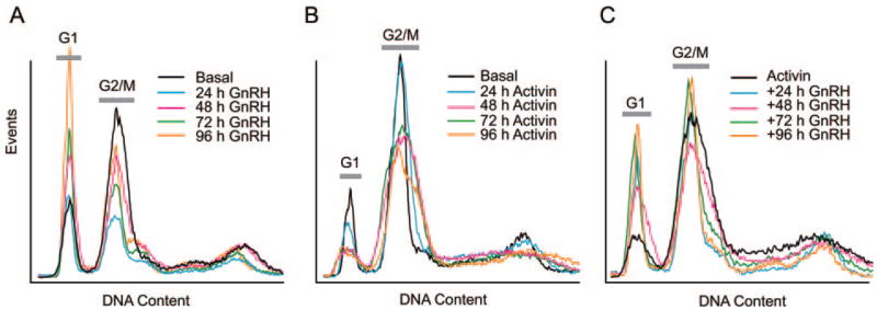
Fig. 11. Effect of GnRH on Cell Cycle by Flow Cytometry.
A, Cells were cultured in complete medium containing 10% fetal calf serum and 100 nM GnRH for increasing times. Cells were fixed in ethanol and stained with propidium iodide. DNA content was measured by flow cytometry. B, Cells were treated with 25 ng/ml activin for increasing times, and then fixed and stained with propidium iodide. C, Effect of activin plus GnRH on cell cycle. Cells were treated with 25 ng/ml activin for 24 h before 100 nM GnRH for increasing times. Cells were fixed and stained. Experiment was repeated three times with similar results. G1 and G2/M peaks are indicated. Horizontal axis shows DNA content; vertical axis shows number of events. Peak at higher DNA content is due to tetraploidy.

