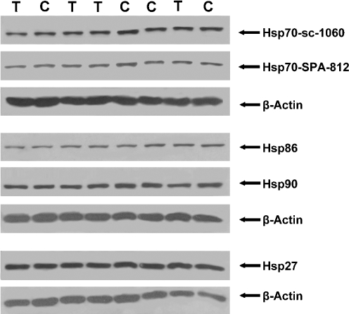
Fig. 3.
Detection of Hsps in the heart of control pigs (C) and 6 h transported pigs (T). Densities of the Hsps in the western blots were normalized to its corresponding heart tissue β-actin as the difference. Here, only a part of the immunoblot that reacted with antibodies specific for Hsp70-sc-1060, Hsp70-SPA-812, Hsp86 (=Hsp90α), Hsp90 (=Hsp90a and βb), and Hsp27 is shown

