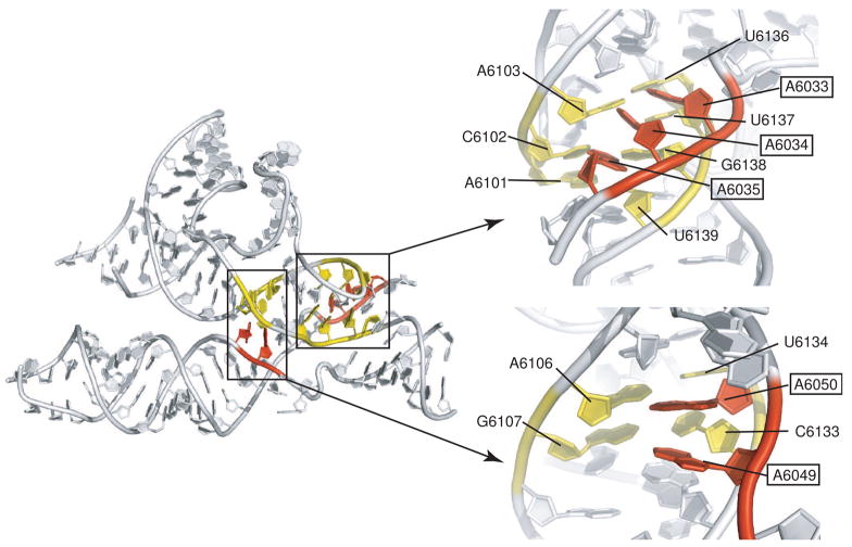
Fig. 4. A-minor interactions between regions 1 and 2 in the PSIV ribosome-binding domain.
At left is the structure of the PSIV IGR IRES ribosome-binding domain, with two locations of A-minor interactions colored and highlighted. At right are close-ups of these two locations presented in an orientation looking into the minor groove of helix P2.2. Nucleotides from region 2 are colored yellow, and adenosines from L1.2 of region 1 are colored red. In both sets of interactions, the Abases contact the minor groove side of the helix. In the interaction at top left, the A-bases are at an angle to the bases of the helix, but contacts are still to the minor groove. In the interactions at lower right, the A-bases are closer to being co-planar with those in the helical stack.

