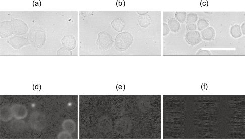
Figure 2.
Bright field (a–c) and photoluminescent (d–f) images of SK-BR-3 cells targeted with PbS-QD/anti-HER2 (~50 nM) (a, d) and PbS-QD/anti-IgG (~50 nM) (b, e) bioconjugates. SK-BR-3 cells incubated with PBS are used as control (c, f). All images are in the same scale with a 50 μm scale bar (40x).
Abbreviations: IgG, immunoglobulin G; PbS, lead sulfide; PBS, phosphate-buffered saline; QD, quantum dots.

