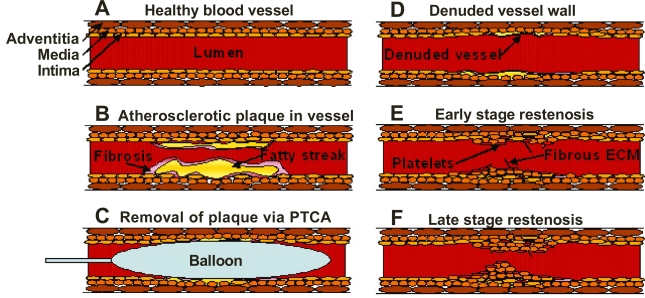
Figure 1.
Schematic illustration of the processes leading to restenotic lesion development. The figures show a healthy blood vessel (A), formation of atherosclerotic plaque within the blood vessel showing a fatty streak and macrophages encapsulated within a fibrotic tissue (B), insertion of a balloon angioplasty catheter to remove the plaque (C), damage due to stripping of the endothelial cells of the vessel wall after removal of the balloon (D), platelet accumulation and activation as well as rapid growth of smooth muscle cells and fibrous extracellular matrix forming the scaffolding (E), and the late stage restenosis showing neointima protruding into the lumen causing occlusion within the vessel (F).

