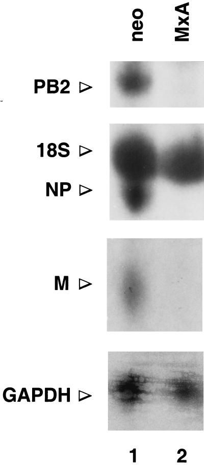
Figure 1.
Detection of primary THOV transcripts by Northern blot analysis. Parallel cultures of MxA-expressing 3T3 cells (MxA, lane 2) or control cells (neo, lane 1) (14) were infected with 20 plaque forming units of THOV (31) per cell in the presence of 50 μg/ml CHX. Total RNA was prepared 7 h after infection, and samples of 20 μg of RNA were loaded into each lane. Northern blots were hybridized with radiolabeled, negative-sense RNA probes derived from the PB2, NP, and M genes of THOV (34), as indicated. For the detection of glyceraldehyde-3-phosphate dehydrogenase (GAPDH) transcripts, blots were analyzed with a specific, nick-translated cDNA probe. Blots were exposed to x-ray films to detect the radioactive signals. The position of the cross-hybridizing 18S rRNA is indicated.

