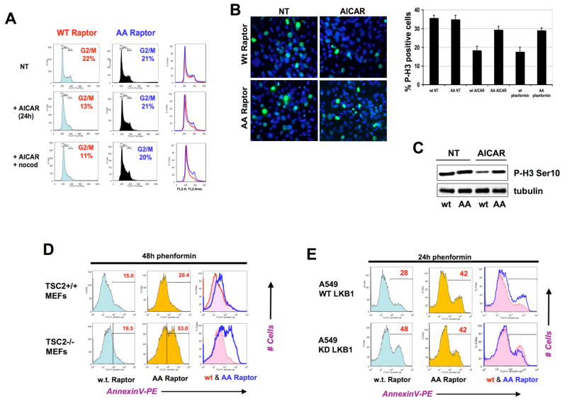
Figure 6. Phosphorylation of Raptor at S722 and S792 dictates a metabolic checkpoint controlling growth arrest and apoptosis in response to energy stress.
A. TSC2−/−, p53−/− MEFs expressing wild-type raptor undergo G1/S arrest following AICAR treatment, while those expressing AA mutant raptor do not. Cells were left untreated (NT) or treated with 2 mM AICAR, or treated with 2 mM AICAR and 3h later exposed to nocodazole (+nocod) to arrest any cycling cells in G2/M. At 24h after AICAR treatment, all cells were fixed and analyzed for DNA content using propidium iodide and FACS analysis. The percentage of cells in the G2/M phase of the cell cycle are highlighted in each population.
B. Cells analogous to those in panel A were plated on coverslips and the next day left untreated (NT) or treated with 2mM AICAR and fixed 18h later. Cells were processed for phospho-histone H3 Ser10 immunocytochemistry to visualize the cells actively going through mitosis at the time the cells were fixed. DAPI was used as a nuclear counterstain. Histogram quantifies Phospho-Histone H3 immunocytochemistry on indicated cells treated with 5mM phenformin, or 2mM AICAR for 18h. At least 300 cells were scored for each condition.
C. Cell extracts from parallel plates to the ones analyzed in panel A were immunoblotted for phospho-histone H3 Ser10 as a marker of the percentage of cells in mitosis.
D. TSC2+/+, p53−/− MEFs or TSC2−/−, p53−/− MEFs expressing AA mutant raptor undergo apoptosis to a greater extent than those expressing WT raptor at later timepoints following energy stress treatment. Cell populations of indicated genotypes were treated with 5mM phenformin and at 48h the percentage of cells undergoing apoptosis was quantified using AnnexinV staining and FACS analysis. Histograms of cells expressing wild-type raptor (red trace) and AA raptor (blue trace) are overlaid in the right-most panel. The percentage of apoptotic cells in the annexin V-positive population is indicated in red at the upper right hand corner of each histogram.
E. Upstream AMPK signals from LKB1 are needed for the protective effect of WT raptor on apoptosis following energy stress. A549 human lung adenocarcinoma cells, which are null for LKB1, were stably reconstituted with wild-type LKB1 (WT) or mutant kinase dead K78I (KD) LKB1 expressing retroviruses. These cells were subsequently stably infected with retroviruses expressing wild-type or AA raptor. Each of the four resulting populations were treated with 5mM phenformin and analyzed for apoptosis as above.

