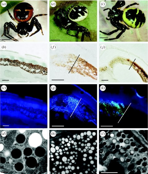
Figure 2.
S. globosum individuals (a–d, e–h and i–l, respectively) of (a) red, (e) white and (i) yellow colours: (a,e,i) habitus, (b,f,j) unstained cross sections of the tegument under light microscopy, (c,g,k) under UV light and (d,h,l) electron micrographs of epithelial cells and pigment granules. The cuticle of both regions, black and coloured (b), is transparent. The absence of fluorescence in the red spider (c) is typical of ommochromes granules (d). In yellow spiders, there is a distinct difference between the black and yellow areas (on the right and left of the dividing mark), both under light microscopy (j) and under UV light (k). The black region contains two types of granules, red and black, whereas the yellow region also contains two types of granules, translucent and light brown (l). Only the yellow portion contains fluorescent granules. In white spiders, the white region (f) contains translucent, fluorescent granules only (g,h). As a result, the white coloration is produced by the guanine layer under the epithelium. Almost the totality of the granules is electron-lucent and homogeneous, indicative of kynurenine (granules type I, Insausti & Casas 2008). There is thus a clear association between body colour and ommochrome metabolites in this non-cryptic crab spider. Scale bars, (a,e,i) 2 mm, (b,c,f,g, j,k) 10 μm, (d) 0.5 μm and (h,l) 2 μm.

