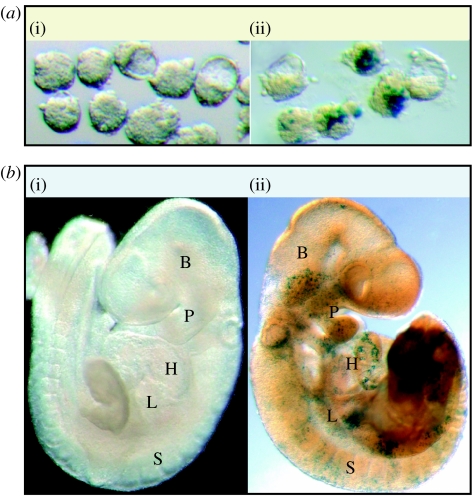
Figure 3.
Mosaic blastocytes derived from labelled embryonic stem cells differentiate as components of multiple organs. (a) 3.5 d.p.c. Mosaic blastocytes: (i) (WT↔WT) normal morphology of blastocyte stage embryos with proper cavitation and inner cell mass formation was observed with (ii) (KO↔WT) lacZ stain highlighting the presence of wild-type embryonic stem cell progeny in the majority of late stage morula: or blastocytes. (b) 9.5 d.p.c. Chimeric blastocytes (i) (WT↔WT) upon intrauterine transplantation and proper development, mosaic blastocytes differentiated into morphologically normal 9.5 d.p.c. with (ii) (KO↔WT) lacZ stain revealing wild-type embryonic stem cell-derived tissue. Expression of lacZ demonstrated wild-type embryonic stem cell-derived progeny throughout the embryo in tissues such as the heart (H), brain (B), somites (S), pharyngeal arches (P) and primordial liver (L).

