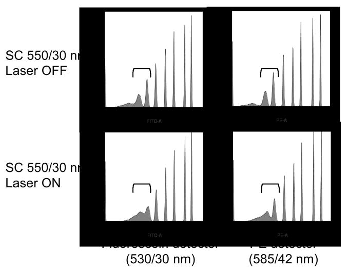
Figure 4.
Spherotech Rainbow premixed eight peak microspheres were analyzed on the LSR II using 488 nm excitation and detection in the fluorescein (530/30 nm) and PE (575/26 nm) detectors, with the SC laser (Fianium) with 550/30 nm filter aligned to an adjacent pinhole and either OFF (top row) or ON (bottom row). Brackets show the resolution between the two dimmest non-blank microsphere populations. The dimmest bead populations are off-scale for all histograms.

