
Figure 1. Over-induction of HIF-1α in HPV31 containing cells.
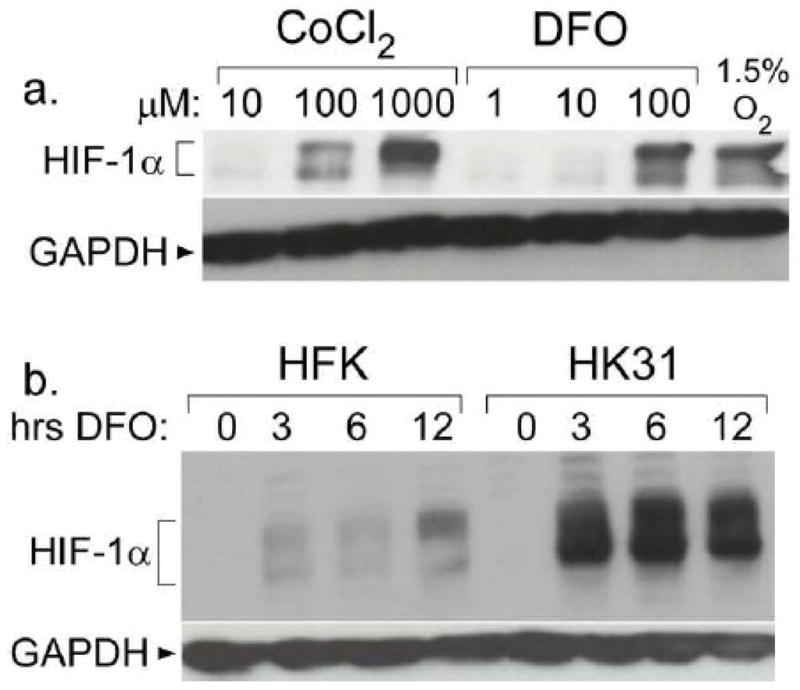
a. CIN 612 cells were treated with the indicated compound for 12 hours or 1.5% oxygen for 48 hours. Total cell extracts were examined for HIF-1α levels by Western blotting. b. HFK-1 and HK31-1 cells were treated with 100 μM DFO for the indicated times, and lysates were analyzed by Western blotting for HIF-1α expression.