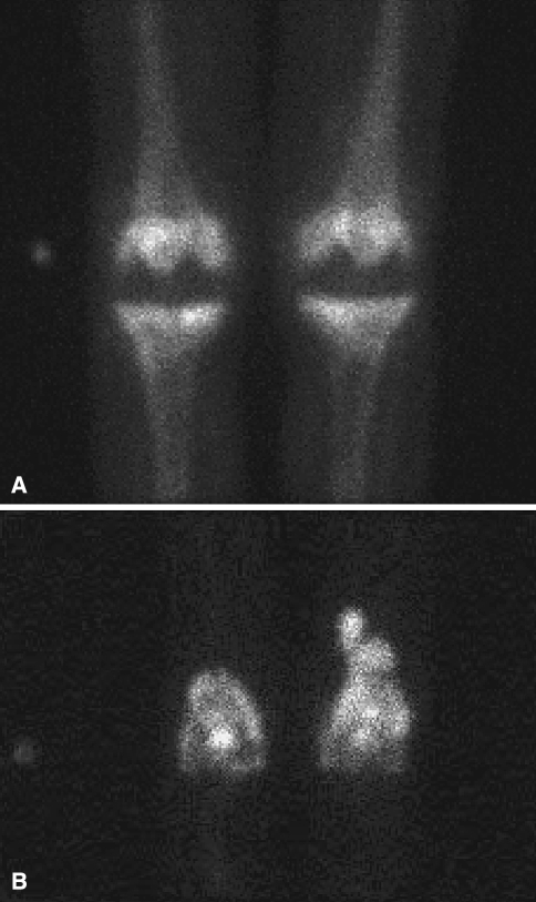
Fig. 2A–B.
(A) A preoperative delayed 3-hour bone scan image shows moderate uptake at the bilateral TKA prostheses. (B) A preoperative indium-labeled leukocyte scan shows discordant areas of increased uptake over the distal femurs, left greater than right, suspicious for bilateral periprosthetic infections.

