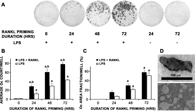
FIG. 1.
Time course study of duration of RANKL priming on P. aeruginosa LPS-mediated Oc differentiation of primary pOcs. pOcs derived from bone marrow cells of C57BL/6 mice were influenced to undergo LPS-mediated Oc formation with media containing LPS and M-CSF with/without RANKL. LPS-mediated Oc formation refers to RANKL (10 ng/ml) priming of pOcs for specified durations prior to P. aeruginosa LPS exposure. A Increases in Oc number were observed with increased duration of RANKL priming. Wells shown here received media containing LPS, M-CSF, and permissive RANKL after priming. B Dose-dependent increases in Oc number were observed with increased duration of RANKL priming in both treatment groups (ap < 0.01, black and white bars). Furthermore, in groups undergoing 24, 48, and 72 h of RANKL priming, Oc formation was always significantly higher in those groups where RANKL was continuously present throughout the experiment (black versus white bars, bp < 0.01). C Time course study of duration of RANKL priming on P. aeruginosa LPS-mediated Oc differentiation of RAW 264.7 cells. RAW cells were influenced to undergo LPS-mediated Oc formation (as above) with media containing LPS with/without RANKL. Dose-dependent increases in Oc area fraction were observed with increased duration of RANKL priming in both treatment groups (ap < 0.01). D Representative photographs of resorption areas formed by primary pOcs on dentin (upper) and hydroxyapatite (lower) during P. aeruginosa LPS-mediated Oc formation. All data are expressed as the mean ± SEM of at least three experiments performed in triplicate wells.

