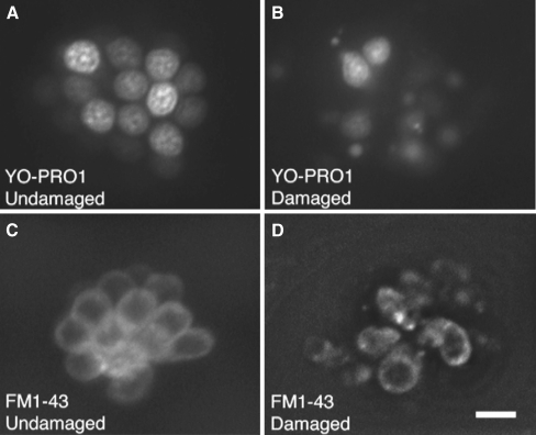
FIG. 1.
Examples of normal and damaged fluorescently labeled hair cells of the zebrafish lateral line. YO-PRO1 selectively labels hair cell nuclei in normal (A) and neomycin-damaged (B) neuromasts. Hair cell protection can thus be easily assessed during screening. For quantitative hair cell counts, FM1-43FX is used to count normal (C) and neomycin-damaged (B) hair cells. In the undamaged neuromast (C) there are approximately 12 visible hair cells. In the damaged neuromast (D), there are two surviving hair cells. Scale bar in D = 10 μM and applies to all panels.

