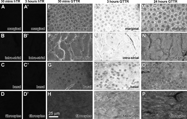
FIG. 3.
Strial uptake and clearance of GTTR. A–D Thirty minutes after i.p. injection of hTR, negligible fluorescence is observed in the different focal planes of a whole-mounted lateral wall (Focal planes determined by corresponding Alexa-488-conjugated phalloidin-labeled image obtained during sequential imaging). A′–D′ Three hours after i.p. injection of hTR, negligible fluorescence is observed in the different focal planes of a whole-mounted lateral wall. E–H GTTR fluorescence in the cytoplasm of marginal cells delineating their nuclei (E), intra-strial tissues (F), basal cells (G), and very weakly in type I fibrocytes (H) 30 min after injection. Note the increased fluorescence in tissues surrounding the strial capillaries in F. I–L Three hours after injection, increased GTTR fluorescence in the cytoplasm of marginal cells, with less intense GTTR fluorescence in intra-strial tissues, basal cells, and fibrocytes in the same focal series. M–P Twenty-four hours after injection, decreased GTTR fluorescence in marginal cells (M), intra-strial tissues (N), basal cells (O) and in spiral ligament type 1 fibrocytes (P) compared to the 3-h time point. All tissues from basal coil of cochlea. Images acquired and post-processed identically.

