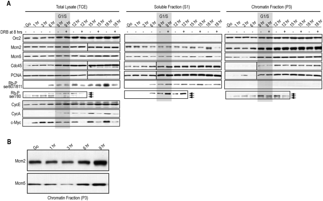
Figure 4. MCM, Cdc45, and PCNA load in the final 3 hrs of G1 in CHO cells.
(A) Parallel to the BrdU and flow cytometry collection in Figure 3 E&F, CHO cells (half treated with DRB at 8 hrs) were collected and separated into total cell lysates (TCE), or fractionated into nucleosolic/cytosolic detergent-soluble extracts (S1) or chromatin-bound detergent-resistant extracts (P3). Immunoblotting with the indicated antibodies was performed on lysates from equal cell numbers loaded into each lane. The G1-S transition in CHO cells (9 hrs after release) is overlayed in gray. (B) An enlargement of the time points from part A for hours G0 through 9 is shown for Mcm2 and Mcm5 immunoblots.

