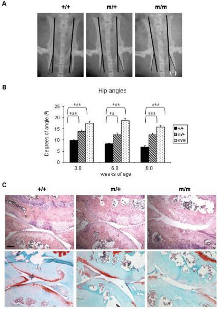
Figure 4.
Mutant mice exhibit a mild hip dysplasia and with age develop severe articular cartilage degeneration. (A) The angle between the tuberosity of the ischium and the pelvic region (°) were measured at 3, 6 and 9 weeks of age. (B) The hip angle was approximately 56% greater for males homozygous for the mutation and 44% greater for males heterozygous for the mutation when compared with their wild-type littermates at 9 weeks (n ≥ 10 mice per genotype; one-way ANOVA). (C) H&E (top panel) and Safranin O (bottom panel) staining of the articular surface of the knee joints at 16 months of age showing severe articular cartilage degradation associated with a complete loss of sulphated proteoglycans in mice homozygous for the mutation. Scale bar 100 μm. Key: +/+ (wild-type); m/+ (heterozygous for the mutation); m/m (homozygous for the mutation); ***P < 0.001; **P < 0.01; *P < 0.05.

