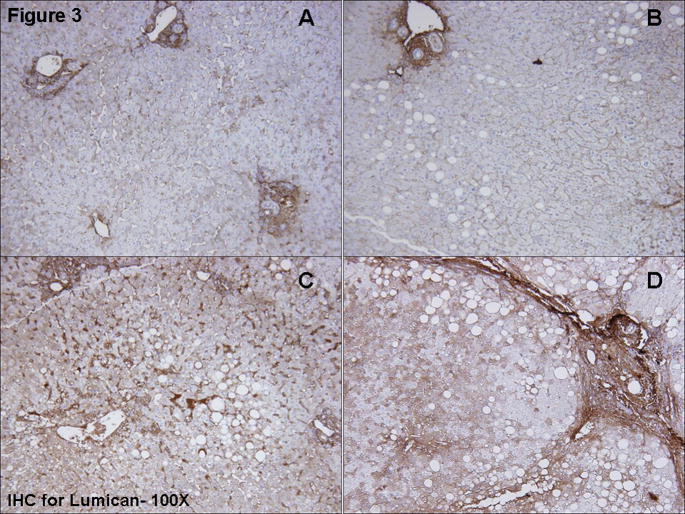
Figure 3.
Immunohistochemical staining for lumican. Moderate stromal staining was seen in portal areas. Staining intensity for lumican in hepatocytes varied between study groups, with more intense staining occurring in NASH-mild (3c) and NASH-severe (3d) than in simple steatosis (3b) or obese normal (3a) biopsies. Hepatocyte staining was cytoplasmic and was apparent throughout the parenchyma but most notable in zones 2–3 in a mosaic pattern. Mild sinusoidal staining was seen. The pattern of lumican staining closely paralleled the fibrosis.

