Fig. 1.
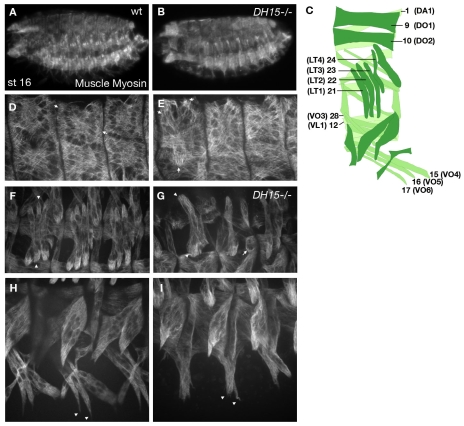
Drosophila RacGAP mutants have defects in somatic muscle patterning. Stage 16 embryos stained for Muscle myosin. Muscle patterning is shown at low (A,B) and high (D-I) magnification in wild-type (wt) (A,D,F,H) or RacGAP50CDH15 (B,E,G,I) embryos. (A) Muscle pattern as observed in the whole embryo. (B) No significant loss of muscle tissue or unfused myoblasts are observed in RacGAP50CDH15 mutants. (D,E) Arrows in D mark normal attachments of DO1. In RacGAP50CDH15, DO1 is in the incorrect position (E, arrows). (F,G) Arrowheads in F mark muscle 22 (LT2). Arrowheads in G mark an LT muscle of an abnormal shape. The arrow marks a rounded muscle that fails to migrate. (H,I) Arrowheads in H mark normal attachments for VO muscles. In RacGAP50CDH15 mutants, these muscles fail to fully extend (I, arrowheads). (C) Schematic of the wild-type muscle pattern of a single abdominal hemisegment, showing the names and numbers for muscles as referred to throughout this work. DA, dorsal acute; DO, dorsal oblique; LT, lateral transverse; VL, ventral lateral; VO, ventral oblique.

