Fig. 8.
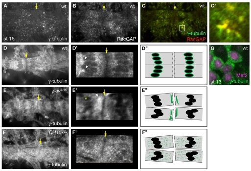
RacGAP controls MT polarity through interaction with γ-tubulin. Yellow arrows mark segment borders, where myotubes from adjacent segments make attachments. (A,B) In wild type, γ-tubulin (A) and RacGAP (B) have a similar localization pattern in cytoplasmic puncta that are concentrated towards the center of the myotube. (C,C′) Merge of RacGAP (red) and γ-tubulin (green) shows that γ-tubulin colocalizes with RacGAP puncta at the nuclear periphery. (D-G) Gain was increased for γ-tubulin imaging in D,D′,E,E′,F,F′,G. The segment borders indicated by the yellow arrows in D, E and F are magnified in D′, E′ and F′, respectively. D″, E″ and F″ are schematics of γ-tubulin localization within extending myotubes at a segment border in the wild-type, pav and RacGAP backgrounds, respectively. In wild-type mononucleated myoblasts (G), γ-tubulin (green) is present diffusely throughout the cytoplasm and is absent from the nucleus (magenta, labeled with anti-Mef2). In wild-type myotubes (D,D′), γ-tubulin is found in the cytoplasm (D), with puncta concentrated at the nuclear periphery (D′, arrowheads), but is absent from the muscle ends (yellow arrow). In pavB200 mutants (E,E′), γ-tubulin is not as highly concentrated around nuclei (E′, n), but is localized to patches at muscle ends (E′, arrowhead). In RacGAPDH15 mutants (F,F′), γ-tubulin fails to localize to perinuclear sites and instead is diffusely localized throughout the muscle cytoplasm, similar to its pattern in mononucleated myoblasts (G).

