
Fig. 4.
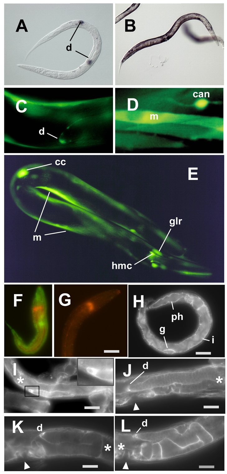
Expression of mig-6 encoded mRNAs and proteins. (A,B) In situ hybridization using a mig-6L-specific probe showing DTC (d) specific expression (A), and using a probe common for mig-6S and mig-6L showing expression in the body wall muscles and other unidentified cells (B). (C-E) From L2 to adult stages, a mig-6 promoter::gfp transcriptional reporter construct is expressed in DTCs (d) (C), in body wall muscles (m), in the CAN neuron (can) (D), in the head mesodermal cell (hmc), in GLR cells (glr) and in coelomocytes (cc) (E). (F,G) Anti-MIG-6 antibody staining of late embryos (released from their shells) shows MIG-6 protein is expressed by body wall muscle primordium (green) in the wild type (F) but not in mig-6(ev788) (G), whereas control antibody, anti-SAD-1 (orange) stains nerve ring and ventral nerve cord in both strains. (H) In early larval stages, anti-MIG-6 antibody stains basement membranes of the gonad primordium (g), pharynx (ph) and intestine (i). This staining persists throughout larval and adult stages. (I,J) Anti-MIG-6 antibody stains basement membrane of the gonad and migrating DTCs (d) of L3 (I) and L4 (J) stages. Enlargement in I shows staining on the DTC. (K,L) The same basement membranes and DTCs are stained by anti-MIG-6 antibody in class-s mig-6(ev700) (K) and in mig-6(k177), which displays a phase 2M DTC defect (L) (see also Fig. S2 in the supplementary material). Vulvae are indicated by white triangles. Asterisks indicate posterior. Dorsal is upwards in all panels. Scale bars: 20 μm.