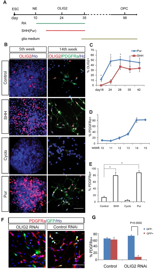
Fig. 1.
SHH-dependent OLIG2 expression for hESC differentiation to OPCs. (A) Experimental paradigm showing differentiation of OLIG2 progenitors and OPCs. (B) OLIG2-expressing progenitors and OLIG2+ PDGFRα+ OPCs are present in the control, SHH-treated and purmorphamine (Pur)-treated cultures, but rarely in the cyclopamine (Cyclo)-treated cultures at day 35 (5th week) and day 98 (14th week). (C) Time course of OLIG2 expression in response to Pur (1 μM) and SHH (100 ng/ml). *P=0.011, 0.008 and 0.014 at day 18, 23 and 28, respectively. (D) PDGFRα+ OPCs first appear after 10 weeks of differentiation and reach a plateau at the fifteenth week. (E) Comparative effect of SHH, purmorphamine and cyclopamine on the efficiency of OPC generation at the fifteenth week. *P<0.0001. The percentage PDGFRα in D and E represents the proportion of PDGFRα+ cells among total cells. (F) OLIG2 RNAi cells do not express PDGFRα (arrows), whereas control RNAi cells are positive for PDGFRα (arrowheads). (G) Quantification of the effect of OLIG2 RNAi on the percentage of PDGFRα+ cells. Cells infected with the control RNAi virus (GFP+) generated a similar proportion of PDGFRα-expressing cells to those without viral infection (GFP-), whereas the cells infected with OLIG2 RNAi virus (GFP+) generated fewer PDGFRα+ OPCs than the non-infected population (GFP-). Ho, Hoechst 33258-stained nuclei. Scale bars: 50 μm.

