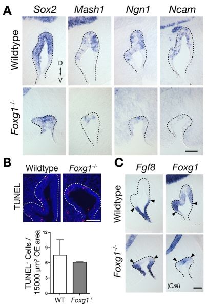
Fig. 1.
Failure of primary neurogenesis in Foxg1-/- OE. (A) Sections of olfactory epithelium (OE) from wild-type and Foxg1-/- mouse embryos at E11, showing the decrease in the numbers of cells expressing stage-specific neuronal markers. D, dorsal; V, ventral. (B) Apoptotic cells visualized by TUNEL labeling in E11 OE from wild-type and Foxg1-/- OE. For comparison, numbers were normalized to an area of 15,000 μm2, the average area of each section of Foxg1-/- OE at this age. Mean values ±s.d. of TUNEL+ cells per 15,000 μm2 OE are: wild type, 7.51±4.24; Foxg1-/-, 6.11±0.095. Data, which showed no significant difference (Student's t-test) (Glantz, 2005), were collected from two animals of each genotype. (C) Fgf8 and Foxg1 expression at E11. Fgf8 is expressed at the rim of the olfactory pit (OP) in wild type, and the pattern is unchanged in Foxg1-/- OE (arrowheads). The Foxg1 expression domain (detected by ISH to Cre), located in the central neurogenic zone of the OE, is reduced in Foxg1-/- OE. Scale bars: 100 μm.

