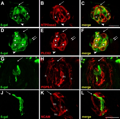
Fig. 4.
Taste placodes give rise to type I and II, but not to type III cells in adult taste buds. Mice treated with tamoxifen at E12.5 were examined for β-gal (green) and differentiated taste cell marker (red) immunoreactivity 6 weeks postnatally. (A-C) Single confocal planes of adult tongue cryosections stained with anti-β-gal (green; cytoplasmic) and anti-NTPDase2 (red; membrane associated) showed many double-labeled cells (white asterisks). Intragemmal basal cells were also labeled by anti-β-gal (white arrowheads). (D-F) Confocal single plane image of a cryosection immunostained for β-gal and PLCβ2 (red; cytoplasm) shows numerous co-expressing cells (white asterisks). An intragemmal basal cell (white arrowheads) and perigemmal edge cell (white double arrows) were also β-gal-IR. (G-I) A taste receptor cells (trc) immunopositive for PGP9.5 (red) was not β-gal-IR. Anti-PGP9.5 also labels nerve fibers (n) in and around buds. (J-L) Staining for β-gal (green) and anti-NCAM (red) did not reveal double-labeled taste receptor cells (trc). Anti-NCAM also labels nerve fibers (n) innervating taste buds. The single white arrow in each panel indicates the taste pore. Scale bars: 20 μm.

