Fig. 1.
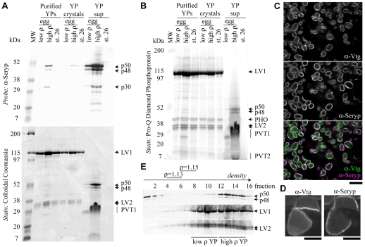
Seryp is a novel component of the YP superficial layer. (A,B) YPs were purified from eggs [low and high density (p) YPs] and tailbud stage embryos (stage 26) and the contents subfractionated into YP crystal and YP supernatant (sup) fractions. Purified YPs and the two derived fractions were analyzed by western blotting (A), Coomassie staining (A) and phosphoprotein staining (B). Seryp species were greatly enriched in the YP supernatant fraction, whereas LV1 and LV2 were nearly exclusively in YP crystals. The phosphoprotein stain identified PHO, PVT1 and PVT2, based on approximate size similarity to the major yolk phosphoproteins described by Wiley and Wallace (Wiley and Wallace, 1981). PVT1 also stained with Coomassie and appeared to migrate anomalously when greatly enriched in the YP supernatant. (C,D) Anti-Vtg-Alexa488 (α-Vtg) and anti-Seryp-Alexa568 (α-Seryp) antibodies colocalize to the rim of YPs in unfertilized egg sections (C). Anti-Vtg-Alexa488 but not anti-Seryp-Alexa568 bound to the interior of YP crystals that cracked during sectioning (D). Scale bars: 10 μm in C; 5 μm in D. (E) When subjected to isopycnic centrifugation, gently lysed single eggs exhibited lower density, low Seryp YPs and higher density, high Seryp YPs, as well as Seryp that did not sediment. Fractions that contained ρ=1.13 and ρ=1.15 density markers are indicated. Smearing on the western blots was likely to be due to Percoll in the gel wells.

