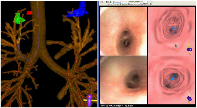
Figure 12.

A screen capture of the guidance system during a live procedure (case 20349.3.28). The left portion shows a global view consisting of an extraluminal rendering of the airway tree, centerlines, and three ROIs (red, green, and blue). The blue centerline depicts the computed route for the green ROI. The yellow cylinder and green protruding needle represent the tip of the virtual bronchoscope at the current viewing site of the procedure. The right portion of the screen capture illustrates endoluminal video (left views) and renderings (right views). The top-left endoluminal rendering represents the current viewing site of the system, as indicated by the virtual bronchoscope, with the blue line indicating the route to follow to the ROI. The top-right view shows the live bronchoscopic video feed. The lower left (endoluminal rendering with blue route) and lower right (bronchoscopic video) views represent a “frozen look” of the previously encountered bifurcation passed while the physician maneuvered the bronchoscope along the route. The physician follows the blue line to reach the ROI. Currently, the physician is 49.8 mm from the ROI, as indicated in the bottom right footer. When the physician reaches the vicinity of the ROI, a view similar to that shown in Figure 7 appears.
