
Figure 5.
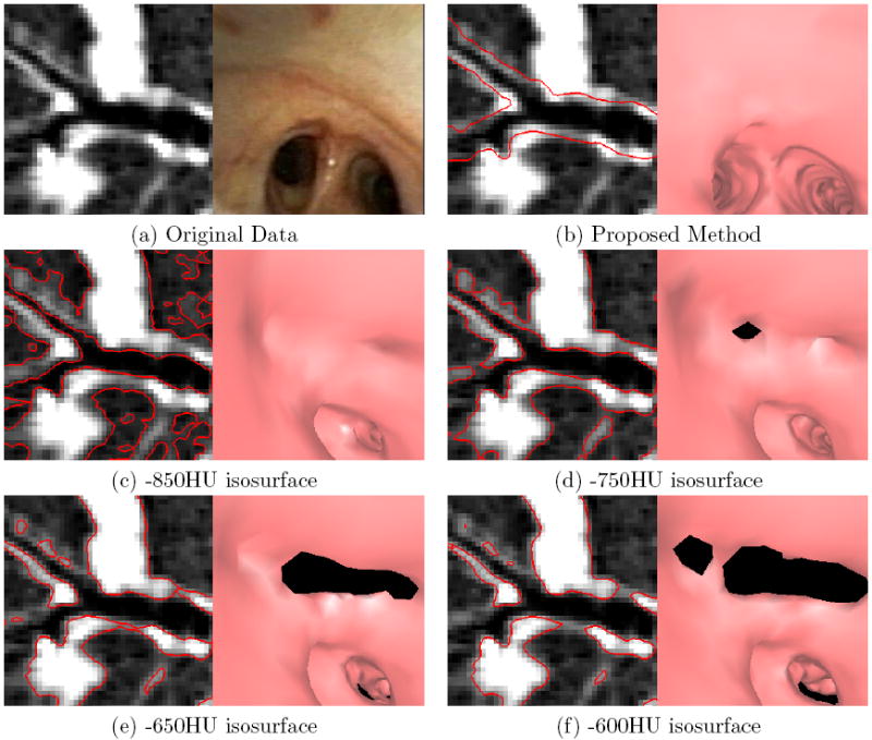
Examples of endoluminal surfaces produced by the proposed surface-definition method and the previous method relying on fixed-HU values. The selected bifurcation is for a peripheral airway near the medial segment of the right upper lobe. A Siemens Sensation 40 scanner generated the MDCT image (image size: 512 × 512 × 557; voxel resolution: Δx = Δy = Δz = 0.50 mm; patient 20349.3.26). (a) Raw local oblique MDCT cross section in the vicinity of the bifurcation and a corresponding bronchoscopic video frame depicting the bifurcation. (b) Surfaces (red) produced by the proposed method overlaid on the cross-sectional image and an endoluminal rendering corresponding to the pose of the real bronchoscopic video of (a). Parts (c-f) depict corresponding results using the fixed-HU method for various HU values. The isosurfaces can be simultaneously too conservatively and too incompletely defined. For all thresholds, the surfaces are too narrow — the bifurcation that is apparent in the video frame (a) is not completely visible in any of the endoluminal renderings (c-f). With the exception of the -850HU isosurface, all isosurfaces also contain noticeable holes (the black regions appearing in the endoluminal renderings).