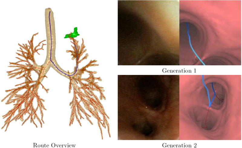
Figure 8.

A bronchoscopic route to a peripheral ROI in patient 20349.3.28. The Route Overview shows a global view of the airway tree, centerlines (red), route (blue) and ROI (green, left upper lobe). The views to the right show video frames taken along the route to the ROI (left) and registered endoluminal renderings of airway surfaces derived by the method of Section 2.3 (right). The endoluminal views are taken along the route at generations 1, the trachea, and 2, the left main bronchus.
