Figure 1.
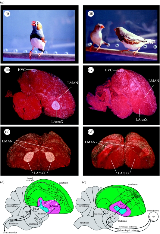
Social context-dependent brain activation differences in the zebra finch and schematics of songbird brain areas involved in singing and vision. (a) Social context difference in the zebra finch brain. (i) Male singing undirected song, (ii) male (zebra-striped chest) singing directed song to a female, (iii) example sagittal section (cresyl violet stained) showing induced egr-1 mRNA (white) in the HVC, LMAN and LAreaX song nuclei after 30 min of undirected singing, (iv) example sagittal section showing the directed singing-driven expression pattern and (v, vi) example frontal sections from other animals showing dramatic differences in LMAN and LAreaX in the same social contexts. (a(i)–(iv)) Modified from Jarvis et al. (1998) and (a(v)(vi)) modified from Hara et al. (2007). (b) Song system: black solid arrows, vocal motor pathway; white arrows, vocal pallial-basal-ganglia-thalamic loop; dashed black arrows, connections between the two vocal pathways; grey arrow, ventral tegmental area dopaminergic projection to LAreaX; weaker projections exist to HVC and RA (not shown). Connectivity of vocal pathways is as summarized in Jarvis (2004); not all connections are shown and only the lateral part of the vocal-basal-ganglia loop is shown. (c) Visual pathways, located lateral to (b): grey arrows, thalamofugal pathway; black arrows, tectofugal pathway; dashed lines, boundary of visual areas as revealed in this study. Connectivity of visual pathways is as summarized in Shimizu & Bowers (1999) and Krutzfeldt & Wild (2004). Green, pallium; pink, striatum; turquoise, pallidum. See abbreviation list for anatomical terms and §2d for further definitions.

