
Fig. 1.
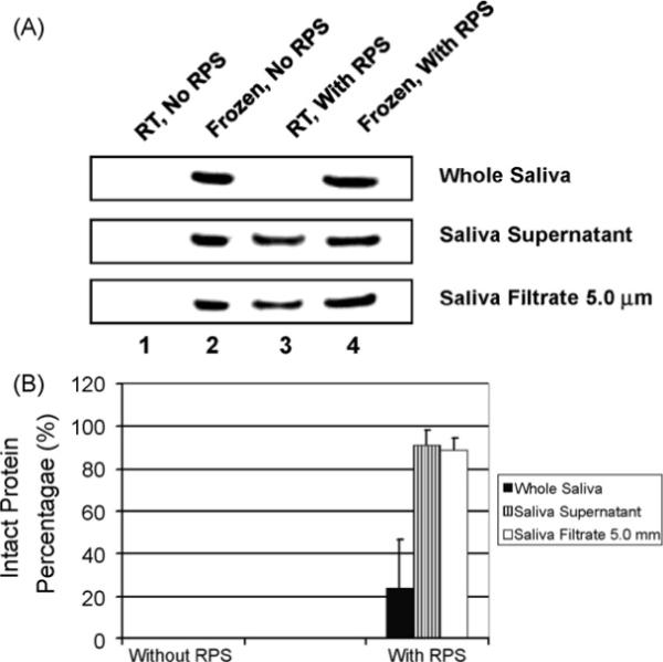
Immunoblotting assay of β-actin after RPS-incubation. (A) Saliva samples, including whole saliva, saliva supernatant and 5.0 μm saliva filtrate were mixed with RPS and incubated at room temperature (RT) for 5 days. After SDS-PAGE, actin was detected by immunoblotting. (B) The intact protein percentage (relative actin level) was calculated by dividing the band intensity of actin in saliva preserved with or without RPS by that of corresponding reference saliva frozen at −80 °C. The error bars represent S.D.