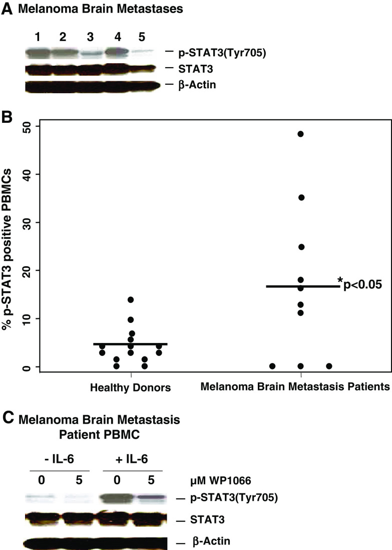
Fig. 1.
Expression of p-STAT3 is enhanced in PBMCs from melanoma brain metastasis patients and is blocked with WP1066. a Melanoma brain metastases express p-STAT3. The resected melanoma metastases were lysed, electrophoretically fractionated in 8% SDS-polyacrylamide gels, transferred to nitrocellulose membranes, and immunoblotted with antibodies to p-STAT3 (Tyr705), STAT3, or β-actin. b Expression of p-STAT3 is enhanced in PBMCs from melanoma brain metastasis patients. PBMCs were isolated from blood samples obtained from healthy donors (n = 14) and melanoma brain metastasis patients (n = 10), fixed in paraformaldehyde, permeabilized, stained with mouse PE-labeled anti-human p-STAT3 (Y705) antibody, and analyzed by FACS. The percentage of p-STAT3-positive PBMCs differed significantly (P < 0.05) between healthy donors and melanoma brain metastasis patients. c WP1066 inhibits p-STAT3 in PBMCs from melanoma brain metastasis patients. The PBMCs were incubated with either the RPMI 1640 medium alone or supplemented with WP1066 in the presence or absence of 10 ng/ml of IL-6. Subsequently, cells were lysed, electrophoretically fractionated in 8% SDS-polyacrylamide gels, transferred to nitrocellulose membranes, and immunoblotted with antibodies to p-STAT3 (Tyr705), STAT3, or β-actin

