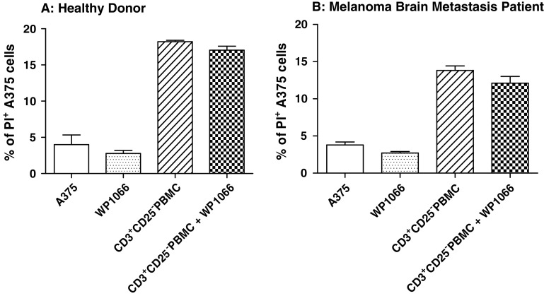
Fig. 4.
Depletion of Tregs eliminates WP1066-enhanced CD3+ T cell cytotoxicity against human melanoma A375 cells. The experiment in Fig. 2 was repeated with CD3+CD25−T cells after depleting Tregs (CD3+CD25+) with a FACSAria cell sorter. A375 cells were labeled with CFSE and exposed to multiple experimental conditions in the presence or absence of CD3+CD25− cells from healthy donors (Fig. 3a, effector:target cell ratio = 3.5:1) and melanoma brain metastasis patients (Fig. 3a, effector:target cell ratio = 10:1). Cells were grown in triplicate cultures in RPMI 1640 medium alone or supplemented with 2 μM of WP1066, CD3+CD25− T cells, or CD3+CD25− T cells plus 2 μM of WP1066. Flow cytometry was used to determine the percentage of PI-positive cells for each experimental condition. *P < 0.05 in comparison with the CD3+CD25− PBMC group. These experiments were reproduced four times with similar results

