
Fig. 6.
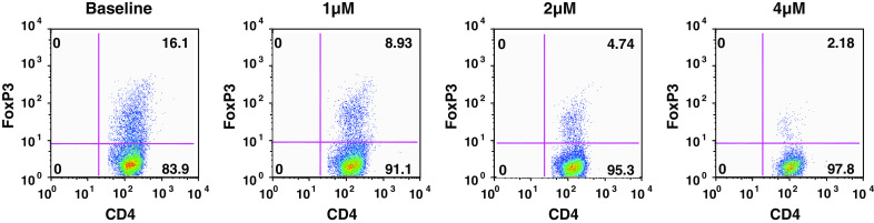
WP1066 inhibits inducible Tregs. CD4+ cells enriched from pooled lymph nodes and spleens of C57BL/6 J mice were stained with PerCP-labeled anti-CD4 (L3T4), FITC-labeled anti-CD62L (MEL-14), and APC-labeled anti-CD25 (PC61) antibodies. A FACSAria cell sorter was used to obtain naïve CD4+CD25− CD62Lhi T cells, which were stimulated with anti-CD3 antibody (2 μg/ml), anti-CD28 antibody (2 μg/ml), and rTGF-β1 (1 ng/ml) to induce FoxP3+ Tregs. The cultures were supplemented with WP1066 at the concentrations shown for 48 h, then stained with PerCP-conjugated anti-CD4 and APC-conjugated anti-CD25 antibodies, permeabilized, and stained with PE-conjugated mAbs to FoxP3. Tregs (high CD4, high FoxP3) are plotted as the percentage of overall T cells for each experimental condition