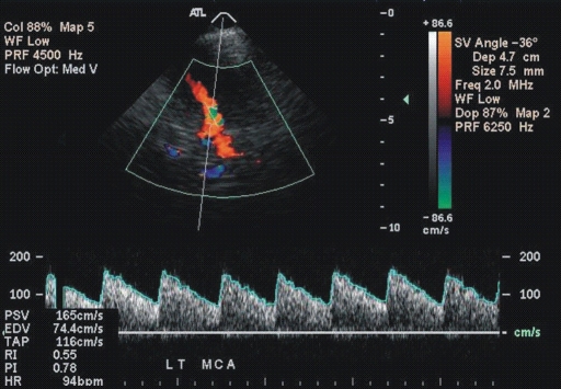
Figure 1a:
(a, b) Transcranial color-coded duplex US scans with typical velocity waveform in a patient with SCD depict (a) left and (b) right MCA in red. (c) Carotid US scan of the left ICA (red). Note that sample volumes are placed on sites of the highest velocity acceleration indicated by aliasing artifacts (green). The angle between the course of an artery and the ultrasound beam indicated by dashed lines is more than 20° (left MCA, 36°; right MCA, 28°), as assumed in the conventional transcranial Doppler US.

