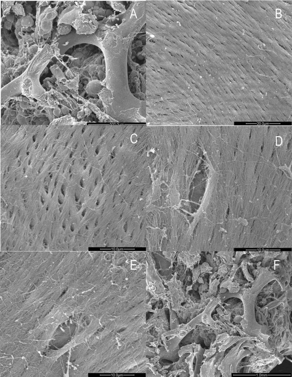
Figure 4.
Images of trabecular bone in osteoarthritic women under SEM. A. Columned trabecular bone of the femoral head in women with OA. (×100). B. Formal collagen fibrils arrayed on the surface of the trabecuae. (×1500). C. Reticular new bone covering the trabeculae. (×3000). D. Some new bones merged. (×3000). E. Lacuna and orderly fibils on the bottom. (×3000). F. Granular new bone tissues inside the trabecular space. (×50).

