Figure 6.
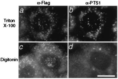
Intracellular localization and topology of Pex19p. N-terminally flag-tagged human Pex19p was expressed in CHO-K1 cells. Cells were treated with 0.1% Triton X-100 (a and b), or with 25 μg/ml of digitonin, under which the plasma membrane was permeabilized (17, 18) (c and d). Cells were stained with antibodies to flag (a and c) and PTS1 (b and d). Note that punctate structures, peroxisomes, are superimposable in a and b and that flag-Pex19p was detected after both types of treatments (a and c). A diffuse staining pattern was partly detected in a and c (see text). (Bar = 20 μm.)

